History
The Pioneering Days of Ultrasound at UW-Madison
In the annals of medical physics at UW-Madison, a remarkable chapter unfolds—the story of ultrasound research. Dr. Charles Kelsey, a nuclear physicist by training, embarked on a journey that would forever alter the landscape of medical imaging at Wisconsin and beyond.
Dr. Charles Kelsey: A Visionary Transition
Dr. Kelsey’s trajectory began with nuclear physics, but destiny nudged him toward a different path. In 1965, he joined the Department of Radiology at UW-Madison, under the mentorship of Dr. John Cameron. The mission? To expand medical physics activities and explore uncharted territories: ultrasound.
Dr. Kelsey and his team—self-described as “tinkerers”—set the stage for groundbreaking research. Within a year, they amassed an eclectic collection of ultrasound devices. From a 2.4 MHz physical therapy unit to a Magnaflux continuous wave Doppler device, their arsenal grew. But skepticism loomed large.
Ultrasound’s Underestimated Potential
In those early days, few grasped the significance of ultrasound. An abstract submitted to the Radiological Society of North America met rejection. The reason? Ultrasound was deemed outside the realm of radiology, its role dismissed. Yet, Dr. Kelsey persisted. You can see an interview of Dr. Kelsey conducted by the very own Dr. John Cameron in 1994 on the AAPM history and heritage website:
Innovations Unleashed
Dr. Kelsey’s graduate student, Jim Zagzebski (who would become chair of the Department of Medical Physics), working with speech researcher Ken Watkin introduced “ultrasonic tongue beads” to study patterns of motion of speed organs. At the time, speech researchers were adhering x-ray-opaque beads to the surface of the tongue. Ken Watkin was hired by Chuck Kelsey and his speech and hearing department colleagues to work on a grant involving use of ultrasound in speech research. The piezoelectric elements used instead of the radiopaque beads adhered temporarily to the tongue surface using a non-toxic dental cementing compound. A single-element transducer, positioned below the chin, emitted ultrasound pulses through the soft tissue. These pulses were detected by the piezoelectric beads after a delay proportional to the transducer-to-bead distance. Dynamic movement patterns of the piezoelectric element on the tongue surface were meticulously tracked using M-mode techniques. By comparing the bead’s motion path to tracings recorded via x-ray cine radiography, the accuracy of this method was convincingly demonstrated. The “tongue beads” technique allowed speech studies without subjecting patients to extensive ionizing radiation, eliminating the need for frame-by-frame x-ray measurements. In addition, Jim Zagzebski built a directional Doppler unit to also record vocal fold motion. The work continued mostly independently of Dr Kelsey after he started working intensely on a different project involving the neutrons. You can check the 1973 publication in the Journal of the Acoustical Society of America here:
Ed Cytacki, another graduate student working with Dr. Kelsey, constructed a contact scanner to replace the water bath scanners in use prior to that time. The articulated arm system enabled 2-D scanning and effectively demonstrated to University of Wisconsin officials the potential of ultrasound for imaging soft tissues in the body. This led to the acquisition of an early commercial version of a “contact scanner” by the UW Hospital Radiology Department. The Picker 80 L is a descendent of one of the first articulated arm scanners developed in the US at the University of Colorado and originally marketed by the Physionics Corporation. Picker purchased Physionics in 1967 and slightly changed the design of the system.
The unit was located in shared Medical Physics –Nuclear Medicine space in the Bradley building attached to the UW Hospital. Initially it was operated by Professor Kelsey and graduate student Jim Zagzebski. Later they trained x-ray and nuclear medicine technician Jackie Cassidy to do scans, and she became UW’s first full time ultrasonographer in the department of Radiology.
The unit used an exchangeable, single element piezoelectric transducer attached to the short arm of the 3-armed tracking device. The angle of each arm was tracked by a series of wires and pulleys attached to a set of sine-cosine potentiometers in the scan console. This provided information on the x-y location and the exact orientation of the single-beam transducer which was operated in a pulse-echo mode. Images were formed on a bistable storage oscilloscope screen, and hardcopy was obtained by photography.
Speech researchers also became fascinated with the 2-D scanning enabled by the articulated arm system. One of the potential applications was in scanning the human tongue. If the transducer is coupled to the skin surface at locations beneath the chin and aimed upward towards the top of the head, the tissue-to-air interface at the dorsal tongue surface provides a strong echo. Thus, it became possible to outline the posture of the tongue during static speech sounds by scanning the dorsal surface. The speaker’s face could be outlined by simply translating the face of the ultrasound transducer over skin with the system sensitivity lowered so that no echoes were displayed. With such a system, the configuration of this important speech organ during various utterances could be brought out.
Ultrasound in Treatment Planning
Ultrasound became indispensable in radiation therapy planning. Depth dose curves overlaid on B-mode images guided tumor localization. The Unirad Ultrasound scanner provided stability and precision. However, the rise of X-ray CT and MRI eventually diminished ultrasound’s role.
Dr. Kelsey’s legacy extended beyond physics. His vision, coupled with the ingenuity of Dr. Jim Zagzebski, shaped a new era—one where ultrasound illuminated the hidden contours of life.
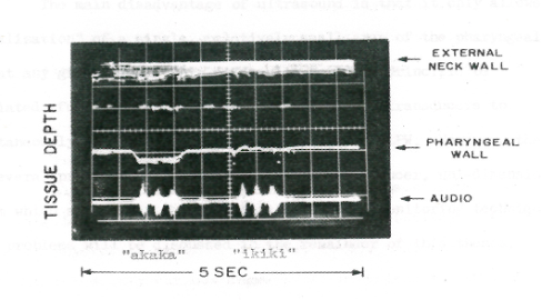
M-mode record of lateral pharyngeal wall motion while the subject uttered “Akaka” and “ikiki,” sounds.
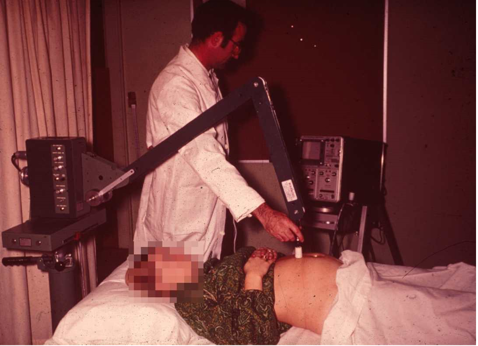
Picker 80 L scanner operated by Professor Kelsey to image a third trimester fetus (~1971).
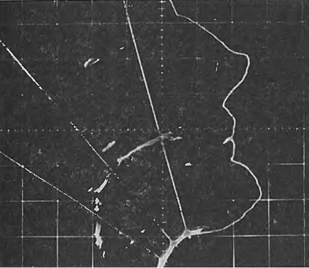
Ultrasound image of the dorsal surface of the tongue while at rest.
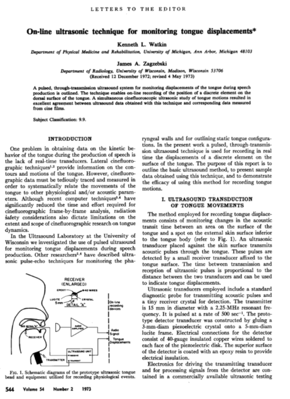
1973 publication by Ken Watkin and Jim Zagzebski on the use of ultrasound to monitor tongue displacement during speech.
Advancements in Tissue-Mimicking Phantoms: A Scientific Endeavor
Within the research corridors of the University of Wisconsin-Madison, a symphony unfolded—a harmonious blend of physics, creativity, and unwavering pursuit. Dr. Charles Kelsey’s legacy had taken root, stretching beyond the visible.
The Quest for Phantom Materials
Diagnostic ultrasound practitioners hungered for tools to assess the capabilities of their devices. The American Institute of Ultrasound in Medicine’s Standard 100 mm test object stood sentinel—a matrix of stainless steel rods, silently beckoning exploration. Yet, the water-alcohol mixture the rods were immersed in lacked speckle-generating properties. It whispered no echoes, painted no contours. Researchers yearned for more—a material that would faithfully mimic the scattering and speckle-generating essence of biological tissue.
Ernie Madsen: The phantom master
In 1976, physicist Ernest Madsen stepped into the spotlight. Ernie’s academic lineage traced back to the University of Maryland and the Catholic University of America. His solid-state background, honed through teaching and research, infused fresh vigor. Ernie’s previous tenure at Radiation Measurements, Inc., where he crafted phantoms for testing x-ray mammography equipment, now intersected with UW-Madison. Dr. Cameron, ever the visionary, established a post-doc position for Ernie within the medical physics group.
Graphite-In-Gel: The Eureka Moment
Ernie’s canvas expanded. Working with Gary Frank, Ernie delved into water-based gels, infusing them with graphite powder—a touch of scientific alchemy. The goal? To discover materials with attenuation coefficients that matched harmoniously with ultrasound frequencies, faithfully mimicking soft tissues. The sweet spot lay between 0.3 and 1.2 dB/cm-MHz. The emergence of graphite-in-gel materials, patent-protected by the Wisconsin Alumni Research Foundation, marked a pivotal moment. Radiology Measurements, Inc. (RMI), the torchbearer, meticulously crafted test phantoms following defined procedures.
The Symphony Resonates
Quality control phantoms, standing about 20 cm high and 15 cm wide, graced ultrasound scanning facilities worldwide. Their easily penetrated scanning windows allowed sound waves to image their complex structures while providing protection from desiccation. String targets, anechoic cylinders, and meticulously calibrated structures—all orchestrated a symphony of precision, enabling ultrasound-system engineers to fine-tune their instruments and clinicians to evaluate their performance. Through the years, Ernie and Gary Frank continued and expanded their phantom designs, creating the phantom lab that would be come a worldwide reference for ultrasound phantom research and would provide the ultrasound industry, clinical, and academic communities with phantoms for decades to come.
Advancements in Quantitative Ultrasound: A Scientific Odyssey
Within the academic corridors of the University of Wisconsin, physicist Jim Zagzebski embarked on a rigorous exploration. His mission: to unravel the intricate physics and practical applications of medical ultrasound. Armed with a Bachelor’s degree in physics from St. Mary’s College, Jim delved into uncharted territory. Little did he know that this scientific pilgrimage would significantly impact diagnostic imaging.
Ernie Madsen, joined Jim’s quest. Together, they forged a path through the complexities of “ultrasound tissue characterization.” Their research spanned dimensions—scattering properties, system performance evaluation, and the complex physics of acoustic scattering.
Graduate students Mitch Goodsitt, Farhad Jafari, and Tom Burke meticulously validated JJ Faran’s scattering theory for solid spheres. Their experiments with stainless steel spheres laid the groundwork for understanding how ultrasound interacts with biological tissues. Bill Davros extended their exploration to spatially distributed glass spheres, while Mike Insana delved into the intricacies of backscatter phenomena.
Tim Hall, an architect of precision, stepped onto the stage. His mission: to bridge the gap between narrowband and broadband pulse-echo systems to fully characterize backscatter from tissue. Evan Boote adapted Tim’s work for clinical imaging systems, bridging theory and practical implementation.
Lin Xin Yao, an astute researcher, recognized the limitations of existing approaches to be implemented in clinical scanners. He introduced the “reference phantom method,” a pragmatic solution allowing simultaneous estimation of broadband attenuation coefficients and backscatter coefficients. The clinical community welcomed this method: it is the basis of several commercial implementations of quantitative ultrasound.
Ernie and Gary Frank orchestrated an interlaboratory comparison of ultrasound tissue characterization methods. Backscatter, attenuation, and speed danced across research groups. Some methods stood firm, while others faltered. The stage was set—a call for reliable techniques echoed through the scientific community.
Tim Hall, now a seasoned investigator, collaborated with Mike Insana at the Radiology Department of the University of Kansas Medical Center. Their quest led them to explore the “effective scatterer diameter,” a metric revealed through the frequency dependence of acoustic scattering. Additionally, they contributed to early developments in strain elastography imaging methods. Hall later joined the UW-Madison Department of Medical Physics in 2003, where he founded and led the Quantitative Ultrasound Lab.
And so, the echoes reverberate—a symphony of waves, a canvas of tissues, and the promise of deeper understanding.
Credits
Text and figures provided by Jim Zagzebski and Tim Hall, with a little help of Microsoft Copilot (April 14, 2024).
This website is a live historical record that will benefit from input from the community. If you have stories, figures, and/or photos related to the history of ultrasound research in the Departments of Medical Physics and Radiology at UW-Madison that you’d like to share, please reach out to Ivan Rosado-Mendez at rosadomendez@wisc.